Cartoons view of the TATA-binding protein highlights the curved surface of the anti-parallel beta-sheet, and the 4 helices on one side. This is a typical α+β arrangement. TBP is bound to DNA, first shown in cartoons, then a spacefilling view.
This view highlights the pseudo-symmetry of the TBP. The N-terminus - in blue- starts with a long beta strand, then a helix, then 4 more anti-parallel beta strands in blue-green, and a long helix in green finishes the first half. Then it continues into the second half of the protein. In this view highlights the pseudo-symmetrical nature of the protein. The red half (C-terminal) is structurally very similar to the blue half (N-terminal). But they are different in that one binds the TATA end of the DNA.
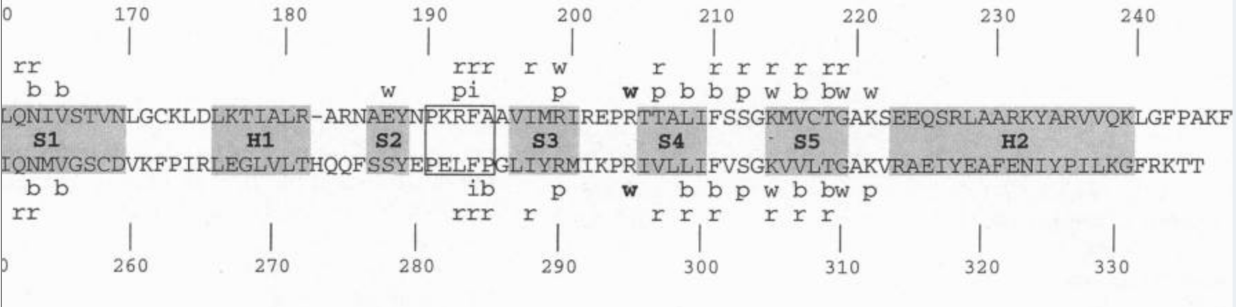
1tgh (PDB)
Juo, Z. S., Chiu, T. K., Leiberman, P. M., Baikalov, I., Berk, A. J., Dickerson, R. E.: How proteins recognize the TATA box. J Mol Biol 261 pp. 239 (1996) (PubMed)
Nikolov DB, Chen H, Halay ED, Hoffman A, Roeder RG, Burley SK.
Proc Natl Acad Sci U S A. 1996 May 14;93(10):4862-7.